
From diagnostic findings to an anatomic map.
Cancer mapping brings imaging, spatially identified pathology and other diagnostic findings together within the anatomy of the prostate.
BioVantra integrates these inputs to estimate the location and extent of disease, preserving the relationship between what was found and where it was found.
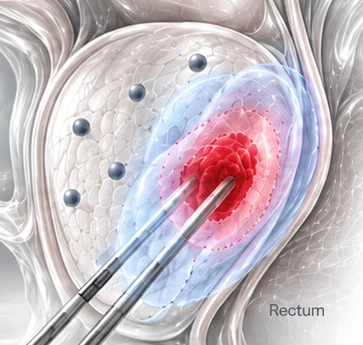
Bringing the findings back together.
Cancer mapping depends on preserving spatial relationships throughout the diagnostic pathway.
Imaging
Establishes the anatomic reference and identifies imaging-defined suspicious disease.
Spatial pathology
Connects diagnosis, grade and measured tumor involvement with the recorded locations from which tissue was obtained.
Cytomolecular findings
Add cellular and molecular observations from defined regions of the prostate when available.
Read together, these findings provide more information than any one component alone.
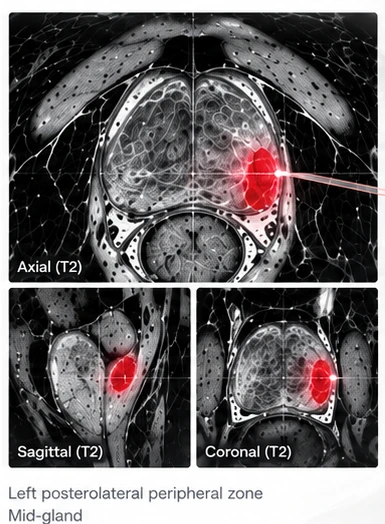
Estimating location and extent.
The integrated map relates diagnostic findings to prostate anatomy to estimate where cancer is present and how far the identified disease may extend.
Imaging-defined lesions provide the initial spatial reference. Biopsy findings establish disease at known sampled locations, while measured tumor involvement and additional spatially identified findings refine the estimate of disease distribution.
The result is an anatomic representation of the estimated cancer volume—not simply a list of positive biopsy sites.
From tumor estimate to treatment margin.
For spatially targeted therapy, the estimated tumor volume can be considered together with a planned treatment margin.
Margin visualization extends beyond the estimated disease boundary to provide an anatomic reference that the treating physician can consider alongside the selected treatment modality, surrounding structures and other clinical information.
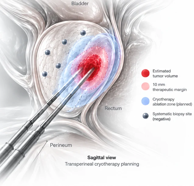
Designed to support spatially targeted treatment planning.
The mapping output is intended to provide the treating physician with an integrated spatial representation of the diagnostic findings.
When focal or targeted therapy is being considered, the map can be reviewed alongside the original imaging, pathology, clinical findings and the technical requirements of the selected treatment.
Preserving the connection between diagnosis and anatomy helps translate diagnostic information into a spatial framework for treatment planning.