
BioVantra — home
Menu
(352) 861-2296 Client Portal

Extending the diagnostic field.

Cytomolecular testing complements core histopathology by extending cellular and molecular sampling across defined regions of the prostate.

When pre-biopsy clinical and MRI findings suggest that focal or targeted therapy may be considered, fine-needle aspiration can be performed alongside core biopsy to provide additional information about the presence, biology and distribution of prostate cancer.

Fine-needle aspiration provides cellular material for cytologic evaluation and appropriate molecular testing while preserving the identity of the region from which each specimen was obtained.

Broader sampling. Additional context.

Core biopsy and fine-needle aspiration provide different but complementary forms of diagnostic information.

Core biopsy

Preserves tissue architecture for histopathologic diagnosis, Gleason grading and quantitative assessment.

Fine-needle aspiration

Can extend sampling across additional defined regions of the prostate without requiring a corresponding core from every location.
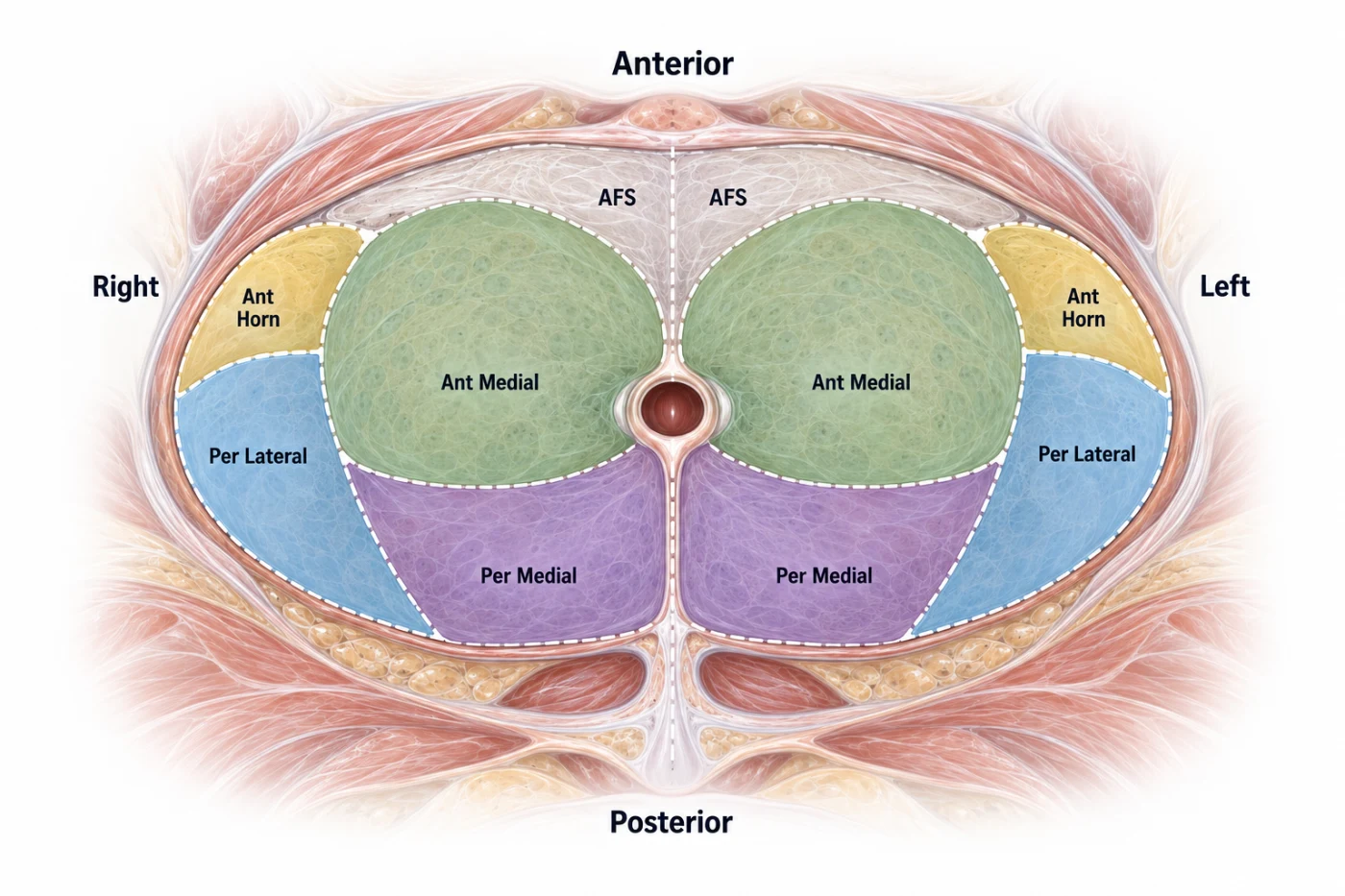
Axial diagram of the prostate showing four defined sampling regions on each side — anterior horn, anterior medial, peripheral lateral and peripheral medial — with the anterior fibromuscular stroma labeled as an anatomic reference.
Eight-region cytomolecular sampling framework. Four defined regions on each side are sampled longitudinally from apex to base; AFS is shown as an anatomic reference.

BioVantra's cytomolecular approach typically samples eight longitudinal regions extending from apex to base: peripheral medial, peripheral lateral, anterior medial and anterior horn on the right, with the corresponding four regions on the left.

Right

Peripheral medial
Peripheral lateral
Anterior medial
Anterior horn

Left

Peripheral medial
Peripheral lateral
Anterior medial
Anterior horn

Multiple aspiration passes within each region broaden the sampled field while maintaining the spatial identity of the specimen.

The objective is not to duplicate the core biopsy, but to add cellular observations from a broader spatial distribution of the gland.
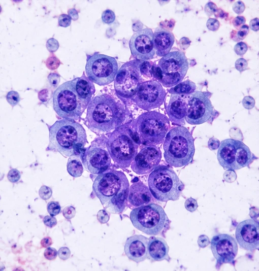
Prostate fine-needle aspiration cytology: a cohesive cluster of malignant epithelial cells with enlarged nuclei and prominent nucleoli, with dispersed cells in the surrounding field.
Prostate FNA cytology demonstrating prostatic carcinoma.

Cellular characterization.

Cytologic evaluation examines the cellular features of material obtained from each sampled region. Findings are reported using a standardized diagnostic framework ranging from no cytological evidence of malignancy through atypical and suspicious findings to prostatic carcinoma.

When carcinoma is identified, cytologic features provide additional information about tumor differentiation while the result remains associated with the region from which the specimen was obtained.

No Cytological Evidence of Malignancy
→
Atypia Without Evidence of Malignancy
→
Atypia Suspicious
→
Prostatic Carcinoma
When carcinoma is identified Low-grade (1/3) · Intermediate (2/3) · High-grade (3/3)
Specimen adequacy Unsatisfactory is reported separately and is not part of this diagnostic progression.

These categories describe cytologic findings in aspirated material and are distinct from Gleason score and Grade Group, which are assigned from histologic evaluation of core biopsy tissue.
Molecular detection and characterization.

Cytomolecular testing can extend beyond cellular morphology to interrogate molecular alterations associated with prostate cancer.

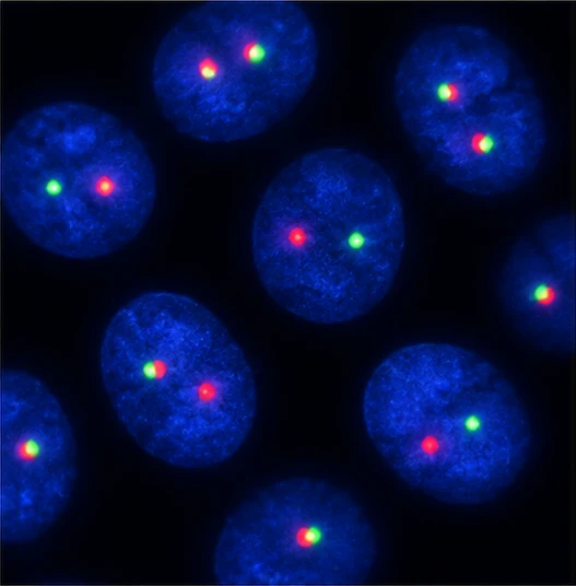
TMPRSS2–ERG FISH evaluates for rearrangement of the TMPRSS2 and ERG genes directly within sampled cells. The rearrangement is identified in approximately half of prostate cancers and is strongly associated with malignant prostatic epithelium. When detected, it can provide molecular evidence supporting the presence of prostate cancer at the sampled location.

The rearrangement pattern also provides an additional dimension of tumor characterization. Because many prostate cancers do not harbor TMPRSS2–ERG rearrangement, a negative result does not exclude malignancy.

TMPRSS2–ERG FISH: interphase nuclei with DAPI counterstain showing separated red and green signals, consistent with a positive rearrangement.
TMPRSS2–ERG FISH demonstrating a positive rearrangement.

Defining disease distribution.

Each cytologic and molecular result remains associated with the region from which the specimen was obtained. Across the eight-region sampling framework, these observations provide additional information about disease distribution beyond the dominant imaging target.

Positive findings can identify additional sampled sites of disease, while negative findings provide complementary information from the regions examined. As with any sampling strategy, a negative result applies to the sampled material and does not establish the absence of cancer throughout the surrounding region.

Informing targeted management.

Disease distribution becomes particularly important when management is intended to be spatially selective.

When focal or targeted therapy is being considered, cytomolecular findings from additional prostate regions can be evaluated alongside MRI and core histopathology to help characterize disease within and beyond the dominant target. The same spatial framework can provide defined regions for subsequent targeted evaluation when surveillance is selected.

Understanding the dominant tumor in the context of the surrounding gland can provide additional information for spatially targeted management.