Prostate biopsy interpretation with preserved spatial orientation.
BioVantra combines subspecialty prostate histopathology with a specimen workflow that preserves each core's identity and orientation from collection through microscopic evaluation.
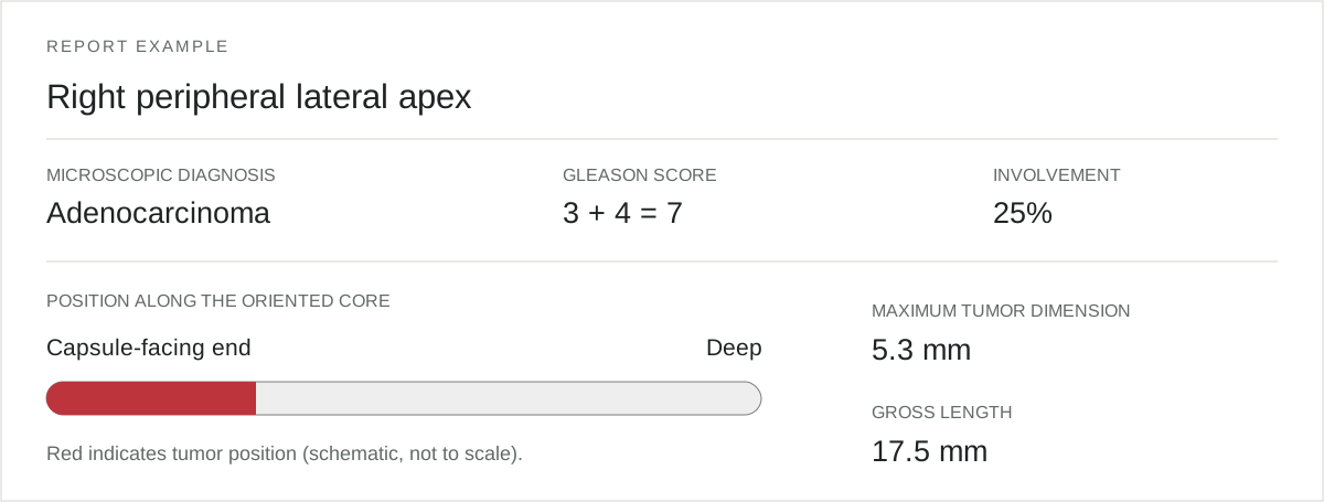
The resulting report connects diagnosis, grade, and tumor extent with the recorded sampling site and tumor position along the oriented core—providing tissue information for subsequent cancer mapping.
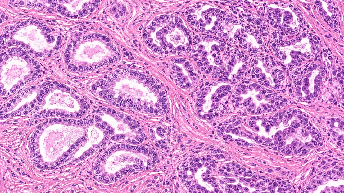
Prostate tissue architecture.
Collection with preserved orientation
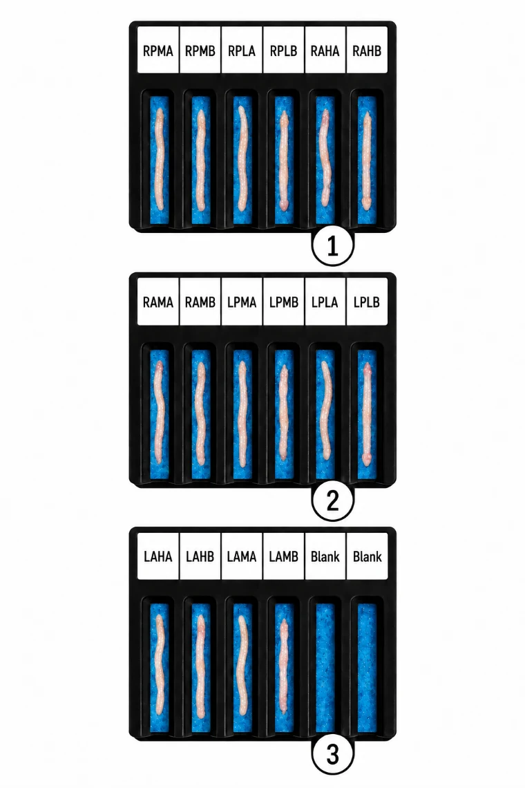
Each biopsy core is transferred directly from the needle into a dedicated, site-identified lane on a BxBoard collection and transport device. The capsule-facing end is identified, and that orientation is preserved throughout laboratory processing.
The BxBoard replaces conventional formalin-filled specimen vials with an organized collection system using a formalin-soaked sponge. Orderly placement helps maintain the specimen's configuration during transport while retaining its connection to the documented biopsy location.
Processing and digitization
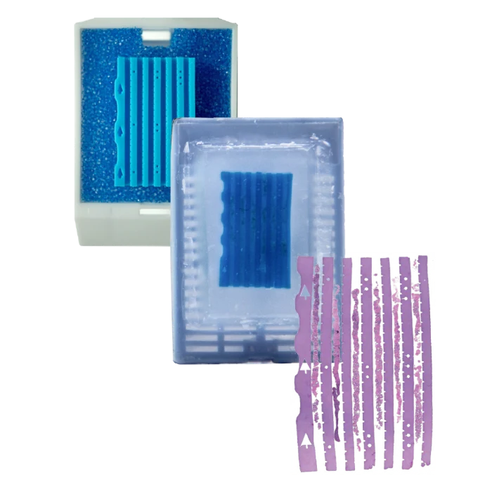
In the laboratory, the cores are transferred into BxChips and photographed for gross examination and specimen measurements.
The BxChips maintain the organized arrangement of the cores through processing, embedding, and sectioning, while the workflow preserves their site identification and capsule-facing orientation. Finished histology slides are digitized for microscopic evaluation.
Gross photographs document the collected tissue before processing. Digitized slides provide the microscopic detail used to evaluate and document the findings.
Subspecialty review with AI assistance
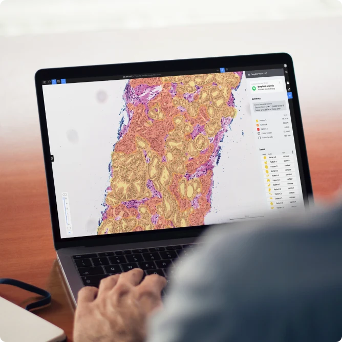
BioVantra's digital pathology workflow incorporates Deep Bio's DeepDx® Prostate to support the pathologist's review of digitized prostate biopsy slides.
The pathologist integrates microscopic findings and supporting analyses, establishes the diagnosis, and signs out the final report.
Diagnosis in anatomic context
- Site-specific diagnosis, grade, and tumor measurements.
- Oriented core and prostate diagrams relating findings to location.
- A patient-focused summary, with ancillary testing status when applicable.
The BioVantra report brings together diagnosis, Gleason score and Grade Group, tumor extent, and findings for each sampled site. Preserved core orientation relates tumor position to the identified capsule-facing end.
Combined with recorded biopsy locations and imaging, these findings contribute to cancer mapping and support discussion between clinician and patient.